Case D701
Many of the lesions on which Cansema
is applied are small epitheliomas (less than 6 mm in diameter,
or under a quarter inch). However, quite a number of cases
have involved skin cancers of considerably larger size.
Quite often these involve life-saving situations, or, in this case,
the prevention of a physician-recommended amputation. Case History: In August, 1991, Mr F.H. of Prineville,
Oregon, wrote our offices (then in Watersmeet, Michigan) and
indicated that he had a serious problem: his upper arm was
heavily diseased with squamous cell carcinoma and the doctors
recommended immediate amputation of the entire right arm
and parts of the shoulder...
![]() Mr. F.H.
indicated that his funds were depleted, and he was
asking if there was anything that we could do to help his situation.
He had read one of our ads in The Spotlight weekly newspaper
and was, therefore, aware of the product. (At that time our
organization went by the name Lenex Laboratories, and Cansema
carried the
brand name Herbveil 8; however, for purposes of clarity, we
shall hereinafter refer to the product given the subject as
Cansema--identical to product we now sell under the latter
brand name.)
Mr. F.H.
indicated that his funds were depleted, and he was
asking if there was anything that we could do to help his situation.
He had read one of our ads in The Spotlight weekly newspaper
and was, therefore, aware of the product. (At that time our
organization went by the name Lenex Laboratories, and Cansema
carried the
brand name Herbveil 8; however, for purposes of clarity, we
shall hereinafter refer to the product given the subject as
Cansema--identical to product we now sell under the latter
brand name.)
![]() We agreed to
supply Cansema on a "pro bono" basis (without charge,
"for free") in return for regular reports and a "photo history"
of Mr. F.H.'s results. After receiving Cansema on September 1st,
Mr. F.H. self-administered Cansema to the lesion on his arm
and began to monitor his results.
We agreed to
supply Cansema on a "pro bono" basis (without charge,
"for free") in return for regular reports and a "photo history"
of Mr. F.H.'s results. After receiving Cansema on September 1st,
Mr. F.H. self-administered Cansema to the lesion on his arm
and began to monitor his results.
![]() The six photos below
tell their own story:
The six photos below
tell their own story:
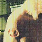
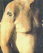
Taken on September 1, 1991, the above photo shows Mr. F.H. before applying Cansema. Notice the deeply pitted area on the upper arm, showing an erosion of the dermis.
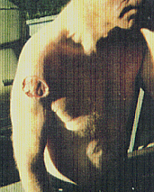
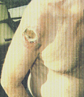
This next picture, taken on September 4, 1991, shows the results of Cansema after 3 days of treatment. There is a slight rise in the affected area ("edema"), and a sharp, distinct definition is already visible between the neoplasm and the healthy surrounding tissue.
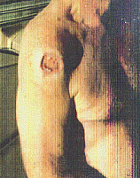
The affected area is a draining heavily, with several changes of bandages required daily. Subject reports considerable pain, though he has elected not to use any analgesics. This photograph shows an eschar forming near the arm pit, despite the fact that Cansema was not applied there. (This is a common phenomenon which we call "transferred necrosis," wherein cancerous cells in the general area are destroyed beyond the initial site of application.)
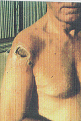
The eschar is continuing to get darker in color, harder, smaller, and more defined. However, the more recently formed eschar near the arm pit has enlarged as it was activated later in the treatment cycle.
Continued heavy drainage. This photo was taken just days before the removal of the eschars. Notice the growth of new healthy tissue around the eschar.
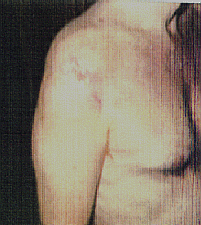
Taken on October 16, 1991, this photo shows a completely healed over skin surface. There is no residual remains of either eschar. The area replacing the carcinoma is tender and a light pink coloring.
The subject has a biopsy of tissue from the affected site to check for residual cancer activity. The result? The testing shows no sign of any remaining cancer.
 Back to Cansema Opening Page
Home Page
Order Form
Back to Cansema Opening Page
Home Page
Order Form