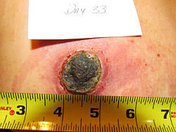
 Update for 2020: Update for 2020: We first posted the pictorial below (and the two cases that follow)
sometime in 1996 -- early in the days of the internet. These cases go back to the early '90s -- our earliest
days at Alpha Omega Labs. The purpose of this page and the two that follow is to show someone who knows little or nothing about
escharotic medicine how the process works and what to expect when you use our Black Salve.
A far more detailed pictorial can be found in Chapter 2 of our book,
Black Salve (2019), or you can view here a pictorial of removal
of skin cancers and keratosis from my own body in Appendix F of that same book
over a 30 year period.  Moreover, hundreds of pictorial examples can be found in the
Cansema testimonial section. [GC]
|
How Cansema Works:
A Pictorial Demonstration

 "In 1989, the incidence of cancer topped one million
for the first time and the number of deaths reached 500,000... (yet)
in the name of orthodoxy, both new and traditional scientific
theories are suppressed, medical records seized, clinics shut
down, and innovative clinicians thrown in prison.
 "But while orthodoxy appears to have all the cards -- money, power,
prestigious credentials, influence in the major media -- the continuing
failure of orthodox medicine to deal satisfactorily with the major
forms of cancer guarantees the growth of nonconventional approaches...
It is the job of the true scientist, and all those who love truth,
to take a serious and open-minded
look at all methods and claims..."
 "A million new cases
a year demand no less." "A million new cases
a year demand no less."
Ralph W. Moss - The Cancer Industry (1989,91)
Pulitzer Prize-Winning Author
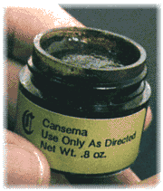
 They say a picture's worth a thousand words. We don't know about
that, but we do know, having worked with thousands of Cansema
users, how our topical formula works. What happens. What are the
stages of cancer necroses and subsequent healing. This we know
quite well.
 So please follow along with us. Below we present you with
three pictorial cases and explain the stages involved, from initial
application through complete healing over of the decavitated area.
-- CASE I --
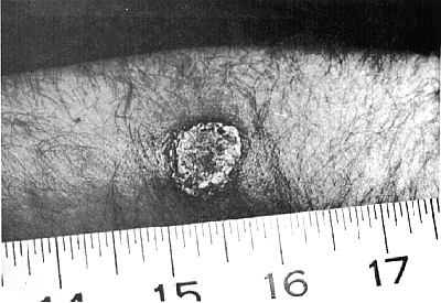
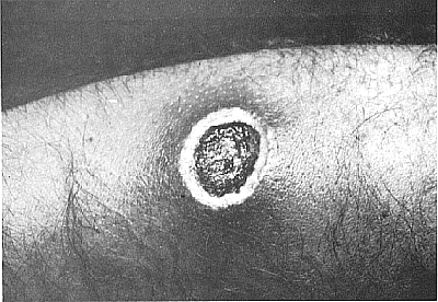
1. ESCHAR FORMATION --
This is the first in a progressive series of photographs taken
following the application of Cansema on a cancerous skin lesion,
this one on the forearm of a middle-aged user from Ohio (USA).
The example above involves one of the more serious types of skin
cancer: melanoma. The photograph above shows that only 30 hours
into the treatment, the entire neoplasm has formed into a scab,
or "eschar." The cancer is completely dead, but the healing
process has only begun.
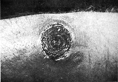
2. EDEMA & ISOLATION --
There is a buildup of antibodies
and serum in the surrounding tissue. As is often typical of edema,
there is a reddening and a general puffiness. The degree can vary
considerably from case to case, but in all ways it is an important
part of the body's healing process. Cansema has successfully
triggered the body's immune system and the necrosis is recognized
as an invasive agent. The eschar becomes better defined from the
surrounding, healthy, non-cancerous tissue.
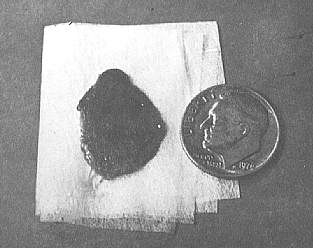
3. ESCHAR CONTAINMENT --
The eschar begins to dry up and contract like any other scab.
As healthy dermal layers are formed beneath the eschar, which nears perfect
and separate formation, it is slowly ejected from the body. We
also call the intermediate step before eschar expulsion "separation."
Edema and redness disappear.
4. ESCHAR EXPULSION --
The entire eschar, representing what had been a thriving
cancer only days before, is pushed out of the body when the last
connective skin tissue beneath it is broken or deteriorates.
What remains at the site of expulsion is a decavitation, which
we will examine next...
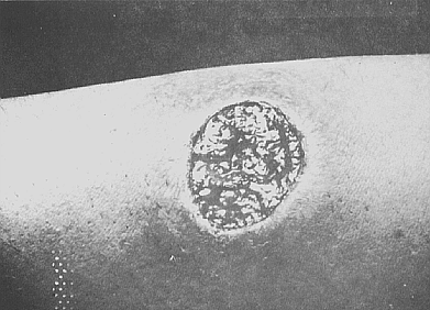
5. DECAVITATION --
Now back at the body... a decavitated area remains where the tumor
was ejected. Epidermal layers have not completely formed, so to the
lay person the area can look extremely raw and unprotected.
Nonetheless, in the thousands of cases we have been involved in,
never once have we had a case of secondary infection resulting
from the process. Vitamin E or petroleum jelly is applied to
minimize scarring and aid the healing process.
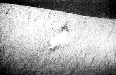
6. FINAL HEALING (also
called "Heal Over") --
The epidermal layers have come in. There is usually minimal scarring
and discoloration, where instructions have been thoroughly followed.
In time even the little scarring seen at right will be marginalized.
 Home Page
 Top of This Page
 Cansema (Top)
 Order Form
|

